AI Oral Lesion Screening: How Vision Models Flag Suspicious Tissue Before Biopsy
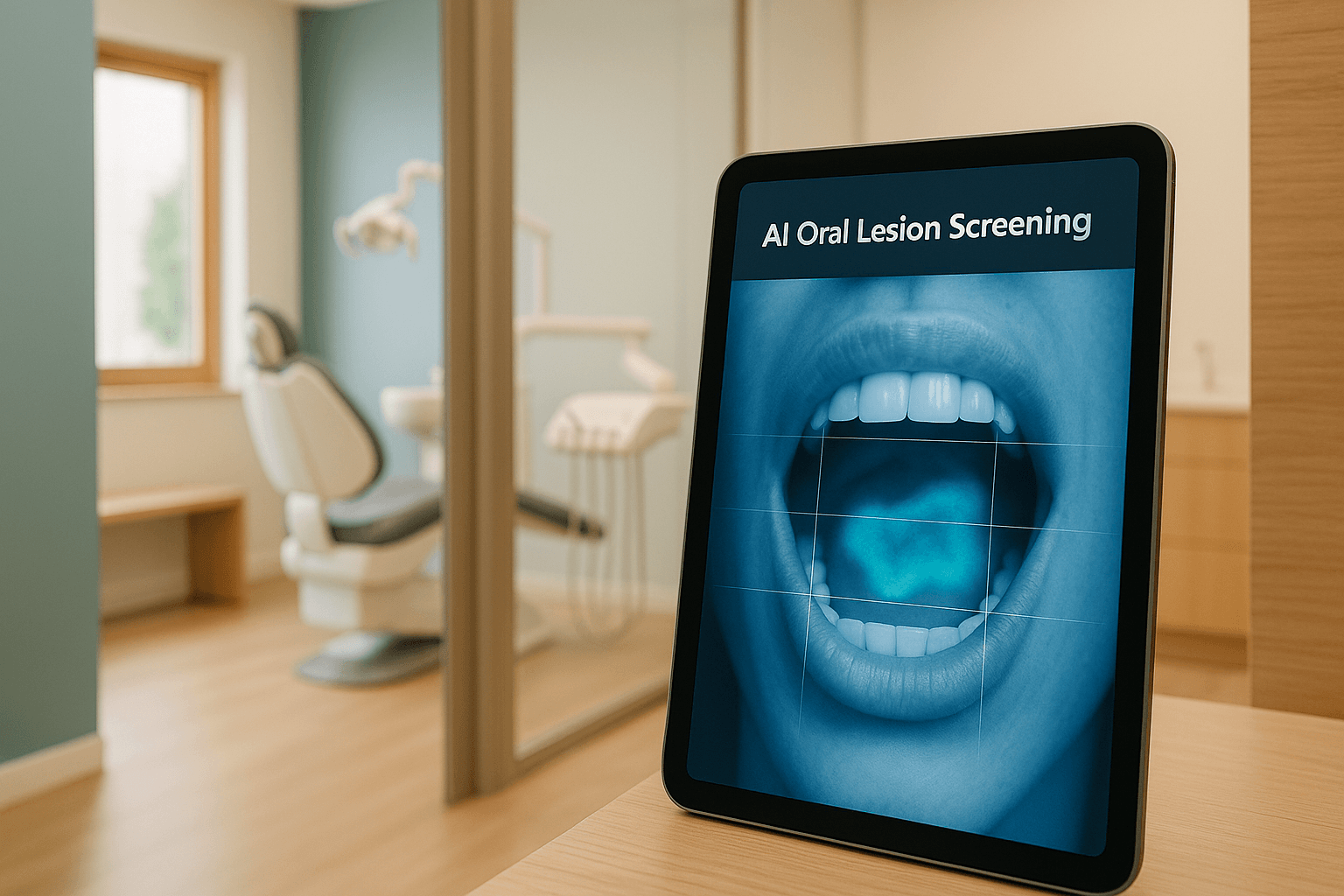
Oral cavity and pharyngeal cancers account for approximately 58,000 new diagnoses and over 12,000 deaths in the United States each year, and the survival gap between an early catch AI airway screening and a late one is enormous. SEER surveillance data puts five-year relative survival above 85 percent for localized disease and near 40 percent once the disease has reached distant sites.
That gap is the entire clinical case for computer-assisted oral lesion screening. It is also the reason this is the one soft-tissue task where a careless deployment does measurable harm.
General dentistry sees more of the mouth, more often, than any other specialty. A hygienist running recare every six months is the highest-frequency observer of oral mucosa anywhere in healthcare, which makes the operatory the right place for a second set of eyes — and the wrong place for anything that sounds like a verdict.
Why Oral Lesion Screening Is Harder Than Caries Detection
Caries detection works because the problem is constrained. A bitewing has fixed anatomy, a grayscale histogram, a canonical view, and a target that presents as radiolucency at a small number of known surfaces, which is why AI caries detection and AI radiograph analysis models generalize across practices as well as they do.
Soft tissue offers none of that. There is no canonical view of the ventrolateral tongue, no fixed anatomy of the buccal mucosa, no grayscale baseline, and an extraordinary range of benign variation that photographs as alarming.
Linea alba, frictional keratosis, geographic tongue, Fordyce granules, amalgam tattoo, physiologic pigmentation, nicotine stomatitis, and reticular lichen planus all present as findings a naive classifier will happily flag. Meanwhile, an early squamous cell carcinoma on the lateral border of the tongue can present as a two-millimeter erythematous patch that photographs as almost nothing.
Then there is the base rate, which is the part most vendors skip in the demo. Malignancy prevalence in an unselected general dental population is low enough that positive predictive value collapses no matter how strong the model is.
Consider the arithmetic on a screen running at 95 percent sensitivity and 90 percent specificity against a population prevalence of roughly 1 in 2,000. That yields about 0.5 true positives per 1,000 patients against roughly 100 false alarms per 1,000 patients — a positive predictive value under one percent, or approximately one real finding per 200 flags.
Keep in mind that this is not a defect in the model; it is a property of screening any low-prevalence condition. It does, however, dictate everything downstream about how output is worded, queued, and staffed.
Oral lesion screening is harder than caries detection because soft tissue has no fixed anatomy, no radiographic baseline, and enormous benign variation. The same white patch can be frictional keratosis or carcinoma in situ.
What Current Vision Models Can Actually Distinguish
Set expectations against what holds up in a real operatory rather than what leads a benchmark table. Published oral lesion classifiers commonly report AUCs in the high 0.80s to low 0.90s on curated datasets, and just as commonly shed ten to twenty points of sensitivity when validated on images from a site that contributed no training data.
What survives that external-validation drop is narrower than the marketing suggests, but it is genuinely useful. The capabilities worth deploying include but are not limited to:
- Presence detection. Separating an abnormal mucosal region from normal mucosa is the model's strongest task and the one that degrades least across sites. This is where the recall you are paying for actually lives.
- Boundary segmentation. Delineating lesion margins and producing a reproducible measurement in millimeters removes the single largest source of variance in serial documentation. Two hygienists eyeballing "about 5 mm" six months apart do not produce comparable data; a segmentation mask does.
- Morphologic class. Sorting a finding into white and keratotic, red and erythroplakic, mixed, ulcerated, pigmented, or exophytic is a reliable and clinically meaningful bucket. Red and mixed lesions carry meaningfully higher risk than homogeneous white ones, and that alone is worth surfacing.
- Anatomic site tagging. Assigning the capture to ventrolateral tongue, floor of mouth, retromolar trigone, soft palate complex, or another region lets you weight risk before any classifier score enters the picture. Site is one of the strongest available priors and it does not require a model to be right about pathology.
- Longitudinal change detection. Registering today's capture against the same site from six and twelve months ago, then quantifying change in area, color, and border character, is the highest-value capability in the entire category. A lesion that is growing, reddening, or losing a defined border is the signal that actually moves a clinician.
Notice that four of those five are measurement tasks rather than diagnostic ones. That is the correct shape for this deployment: the model measures and organizes, and the clinician decides.
Current vision models reliably flag presence, boundary, color class, site, and change over time. They separate visually obvious lesions from normal mucosa well; they do not separate dysplastic from reactive tissue.
The distinction between measurement tasks and diagnostic tasks maps cleanly onto where these systems earn their keep and where they fail:
| Task | Model performance | Correct deployment |
|---|---|---|
| Detect abnormal mucosa vs normal | Strong; degrades least across sites | Automated flag into a clinician review queue |
| Measure lesion area and margin | Strong and highly reproducible | Auto-populate serial measurement in the chart |
| Classify color and surface morphology | Moderate; sensitive to lighting and glare | Risk-weighting input, shown with the image |
| Detect interval change across visits | Strong when capture is standardized | Primary escalation trigger |
| Grade dysplasia | Not possible from surface imaging | Biopsy and histopathology only |
| Distinguish lichen planus from lichenoid dysplasia | Unreliable | Biopsy and histopathology only |
What Vision Models Cannot Distinguish
This section matters more than the previous one, because every serious failure mode in this category comes from a team that read the capability list and quietly assumed the rest. A surface photograph carries a bounded amount of information, and no amount of parameter count adds information the sensor never captured.
The limits that are structural rather than temporary include:
- Dysplasia grade. Mild, moderate, and severe dysplasia are defined by cellular architecture and maturation through the epithelium, which is visible only under histopathology. Two lesions with identical clinical appearance can return hyperkeratosis and carcinoma in situ.
- Lichenoid dysplasia versus lichen planus. These overlap clinically to a degree that defeats surface imaging entirely, and the management difference is significant. This is a biopsy question, permanently.
- Traumatic ulcer versus early carcinoma. Inside the first two weeks these are frequently indistinguishable on a photograph, which is precisely why the two-week persistence rule exists and why a model score cannot substitute for it.
- Submucosal disease. A lesion under intact mucosa may produce almost no surface signal while being clinically obvious to a finger. Cameras do not palpate, and induration, fixation, and cervical lymphadenopathy are outside the model's input space entirely.
- Oropharyngeal sites out of view. HPV-associated disease at the base of tongue or in the tonsillar crypts is often invisible on any intraoral capture. A clean screen is not a clean patient, and the interface should never imply otherwise.
- Anything under blood, saliva, or specular glare. Occluded tissue is not negative tissue, and a model that scores an unusable image is worse than one that refuses it.
Every item on that list is a reason the output has to be framed as suspicion rather than finding. The model narrows attention; it does not close questions.
No vision model can grade dysplasia from a surface photograph. Grading requires cellular architecture visible only under histopathology, so screening output can rank suspicion — never stage disease.
Where The Referral Threshold Belongs
The most common design error in this category is putting the referral threshold on the model's confidence score. That places a low-prevalence classifier in the position of gating a decision whose false-negative cost is a delayed cancer diagnosis.
Put the threshold on persistence instead. Any lesion flagged by the system or noticed by the clinician that remains after two weeks of observation warrants referral or biopsy, regardless of what the score said.
Model confidence still does useful work — it just does it upstream of the decision. Use it to order the review queue, to decide which captures get a second clinician's eyes, and to decide which patients get a shortened recall interval rather than a standard one.
Alongside score, the factors that should weight escalation include:
- Anatomic site. Ventrolateral tongue, floor of mouth, and the soft palate complex carry disproportionate risk and should escalate at a lower bar than buccal mucosa or hard palate.
- Color class. Erythroplakic and mixed lesions escalate above homogeneous leukoplakic ones, which is a well-established clinical prior the software should encode rather than relearn.
- Interval change. Growth, color shift, or border change against a prior capture escalates regardless of absolute score, because trajectory carries more information than any single frame.
- Patient history. Prior oral malignancy, tobacco and alcohol exposure, and immunosuppression change the pretest probability enough that the same image warrants a different disposition.
Escalation is one-directional. A model score may raise a lesion's priority, shorten a recall interval, or trigger a second review. It must never be usable as a reason to skip a referral, and the interface should make de-escalation on model output structurally impossible rather than merely discouraged in training.
That rule is the load-bearing design constraint of the entire deployment. Build it into the state machine, not the policy document, because a policy document does not stop a busy operatory at 4:40 on a Friday.
Set the referral threshold at two weeks of persistence for any flagged lesion, not at a model confidence score. Confidence orders the review queue; persistence and clinical judgment drive the biopsy decision.
Why Screening Output Must Never Be Framed As A Diagnosis
There is a regulatory line here that operators should understand before they write a single string of interface copy. Software that marks a location for clinician attention and software that characterizes what that location is are treated as different categories of device, and the second carries substantially heavier evidentiary and clearance obligations.
Detection-style output — here is a region, review it — sits on the safer side of that line. Characterization-style output — this region is 82 percent likely to be dysplastic — does not, and it also fails clinically for all the reasons in the previous section.
State-level scope-of-practice rules layer on top of the federal question, and they vary considerably; the constraints on what software may present to a patient versus a licensed provider are covered in more depth in our breakdown of state dental board rules for clinical AI. Accordingly, the language rules should be enforced at the template level rather than left to whoever writes the release notes.
The output contract that keeps a screening tool on the right side of that line looks like this:
- No lesion names. The system says "area flagged for clinician evaluation," never "leukoplakia," "lichen planus," or any other clinical entity.
- No malignancy probability in the patient view. A percentage attached to the word cancer is a diagnosis in every way that matters to a patient, regardless of the disclaimer beneath it.
- No treatment or management suggestion. The tool does not recommend biopsy, excision, or observation; it surfaces the image, the measurement, and the change, and the clinician documents the plan.
- Separate surfaces. Clinician-facing detail and patient-facing summary are different renders of the same record, with different permitted vocabulary.
Consent deserves its own attention here, because photographing suspicious tissue and running it through an automated system is a disclosure-worthy step in most practices. Our guide to informed consent for AI-assisted clinical workflows covers the language patterns that hold up, and the same principles that govern AI periodontal screening disclosures apply cleanly to soft-tissue capture.
Screening output must read as a flag for clinician review, not a finding. Label it as an area of interest requiring evaluation, and never emit a lesion name, a malignancy probability, or a treatment suggestion.
The Capture Problem Is Bigger Than The Model Problem
Most oral lesion deployments that underperform do so at the sensor, not the network. Operatory lighting varies in color temperature, saliva produces specular highlights that erase surface texture, retraction is inconsistent, focus is unreliable at close range, and the camera itself changes whenever a practice reorders supplies.
A model trained on cross-polarized captures from a single intraoral camera will not hold its numbers on phone photographs taken under an overhead operatory light. That is domain shift, and it arrives silently.
A capture protocol worth deploying specifies at minimum:
- A fixed view set. Six to nine standardized views covering lateral and ventral tongue, floor of mouth, buccal mucosa bilaterally, hard and soft palate, and labial mucosa, captured in the same order every time.
- Retraction and drying. Consistent retraction and a brief air dry before capture eliminate the two largest sources of unusable frames.
- Cross-polarization. A polarizing filter suppresses specular glare and recovers surface detail that is otherwise lost to wet tissue.
- Fixed working distance and a color reference. Consistent distance makes measurement comparable across visits, and an in-frame color card makes color class comparable across lighting conditions and devices.
- Device registration. Every capture records the camera model and firmware, so a hardware swap is a visible event rather than an unexplained metric drop.
That last item is the one teams skip and later regret. Camera swaps, firmware updates, and operatory light replacements are the most common triggers for the kind of quiet performance decay covered in our piece on model drift monitoring for clinical AI.
One more capture rule earns its complexity: the system should refuse to score an image it cannot use. A reject-and-recapture gate that flags glare, motion blur, occlusion, or off-target framing before inference costs a few seconds — and the chairside latency budget can absorb that far more easily than the chart can absorb a confident score on an unusable frame.
How Do You Know Your Oral Lesion Screening Is Working?
Evaluating this system is harder than evaluating caries detection, because your ground truth is biopsy and you only biopsy what somebody already suspected. That is verification bias, and it means your measured sensitivity is computed on a denominator that excludes exactly the cases you most need to count.
Any honest evaluation has to attack that denominator directly. Note that a validation suite built only on confirmed cases will report excellent numbers on a system that is missing lesions every week.
The metrics that actually tell you something include:
- Interval findings. Any oral malignancy diagnosed within twelve months of a screen the system called clean is the single most important number you track. Instrument the path that lets those come back to you, because they will not arrive on their own.
- Sampled negative follow-up. Pull a random sample of unflagged captures for blinded clinician re-read each quarter, which gives you a defensible estimate of what the queue is missing.
- Per-site recall. Report sensitivity separately for floor of mouth, ventrolateral tongue, and the posterior sites, because aggregate recall hides failure concentrated in the highest-risk regions.
- Per-mucosal-tone subgroup performance. Models trained predominantly on lightly pigmented mucosa underperform on physiologically pigmented tissue, where benign racial pigmentation, amalgam tattoo, and genuine melanotic lesions all compete for the same visual signature. Alert when any subgroup's recall falls more than five points below baseline.
- Alert burden per operatory-day. Flags per hygienist per day is an operational metric that predicts abandonment better than any accuracy figure, and it should be a tracked SLO rather than an afterthought.
Run all of it in shadow mode first. Ninety days of silent inference alongside normal clinical workflow gives you real alert burden, real subgroup numbers, and real disagreement rates before a single flag reaches a clinician — the shadow-mode and validation-suite patterns in our guide to clinical AI evaluation transfer directly to this use case.
Measure sensitivity against biopsied lesions and sampled negatives, not the flagged set alone. Track per-site and per-mucosal-tone recall separately, and alert when any subgroup drops five points from baseline.
What Belongs In The Chart
Everything above only matters if the record can reconstruct what happened. A flagged lesion with no durable trail is a liability rather than a screen.
Each screening event should write, at minimum:
- The image and its capture metadata. Device, firmware, view, timestamp, and any quality-gate result, so a later reviewer knows what the model was looking at.
- The pinned model version. Version and weights hash, because a retrospective review two years later has to reconstruct the exact system that produced the output, not the current one.
- The raw output. Score, segmentation mask, site tag, and morphologic class, stored even when the clinician disagrees.
- The clinician disposition. Agree, disagree, refer, biopsy, or observe, with the override reason captured as structured data rather than free text.
- The follow-up date and its outcome. A two-week re-evaluation that never gets scheduled is the most common way a good screen turns into a bad outcome.
Intraoral photographs tied to a patient record are protected health information without qualification, which puts every processing hop in scope. The architecture requirements — signed BAA, encryption in transit and at rest, access logging on every image read — are covered in our breakdown of HIPAA-compliant clinical AI in dental practices, and vendor diligence for anyone touching those images is covered in SOC 2 evaluation for clinical vendors.
Finally, close the loop operationally. The two-week recheck should generate a scheduled appointment automatically from the flag event, written into the same structured record your AI dental charting workflow already maintains, so persistence gets measured rather than assumed.
Frequently Asked Questions About AI Oral Lesion Screening
The questions below come up in nearly every scoping conversation on this workflow.
Can an AI model tell if an oral lesion is cancerous?
No. Surface photography cannot resolve cellular architecture, so dysplasia grading and malignancy determination require biopsy and histopathology. The model ranks suspicion and prioritizes review; the pathologist makes the call.
What referral threshold should an AI oral screening tool use?
Persistence, not model score. Any flagged lesion that remains after two weeks warrants referral or biopsy regardless of confidence, and the score should only order the review queue rather than gate the decision.
How accurate are oral lesion vision models in real practices?
Curated-dataset AUCs commonly land in the high 0.80s to low 0.90s, but external validation on images from a new site often costs ten to twenty points of sensitivity. Measure on your own captures before trusting either number.
Why do false positives dominate oral cancer screening output?
Base rate. At a prevalence near 1 in 2,000, even 95 percent sensitivity and 90 percent specificity produces a positive predictive value under one percent, or roughly one true finding per 200 flags.
Do intraoral photographs count as PHI under HIPAA?
Yes. Images tied to a patient record are PHI, so any vendor processing them needs a signed BAA, encryption in transit and at rest, and audit logging on every access.
Should AI screening results be shown directly to patients?
Not as a score or a lesion name. Patients should see the photograph and the clinician's documented finding, while raw model output stays on the clinician surface framed as an area flagged for evaluation.
Scoping An Oral Lesion Screening Deployment
Oral lesion screening rewards a narrow, well-instrumented deployment and punishes an ambitious one. The systems that work measure, organize, and escalate; the systems that fail try to characterize.
If you are scoping oral lesion screening and want a second set of eyes before it touches a live operatory, the team at NexV builds and operates HIPAA-grade clinical AI inside dental practices every week. Reach out for a working session — we will review your capture protocol and image quality gate, define the subgroup and interval-finding metrics your evaluation is currently missing, and hand back a shadow-mode plan with escalation language your clinicians can sign off on.