AI Caries Detection: How Algorithms Read Bitewing Radiographs for Early Decay
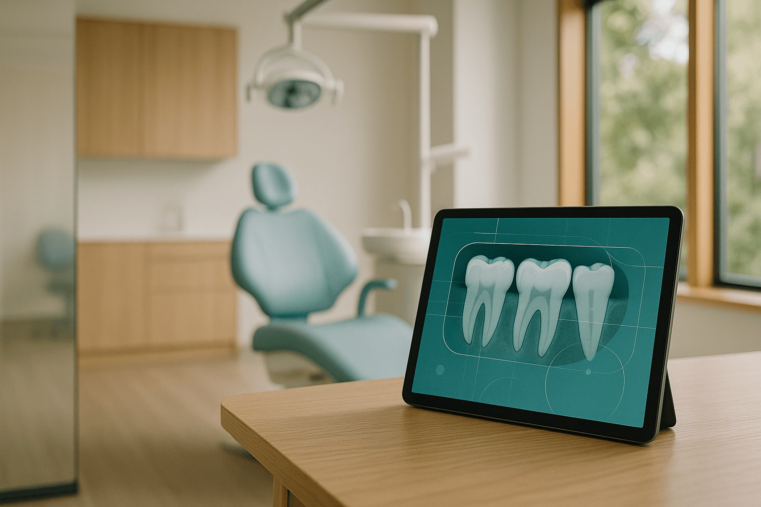
Walk into any dental practice rolling out clinical AI in 2026 and ask which model went into production first. It is almost never perio screening, almost never endo — it is caries detection on bitewings, because that is where the volume lives and where the diagnostic disagreement between clinicians has been documented for thirty years.
Interproximal decay between molars hides in two to four millimeters of overlapping enamel. The same bitewing read by two associates on the same Tuesday produces different treatment plans more often than any operator wants to admit, and that variance is exactly what a well-trained convolutional model is built to compress.
Why Bitewings Are The Highest-Value Surface For Clinical AI
Bitewings are the densest source of diagnostic signal in a routine hygiene visit. Every recall appointment generates two to four of them, every image is captured under near-identical geometry, and every lesion the model surfaces maps to a billable, codable intervention.
That combination — high volume, low geometric variance, and a clean economic loop — is why caries detection is the natural anchor cluster for any clinical-AI rollout. We covered the broader imaging stack in our deep dive on AI radiograph analysis in modern dental practices; this post zooms in on the single pathology where the math works hardest.
How The Algorithm Actually Reads The Image
A production caries model is rarely a single network. It is a small pipeline — preprocessing, tooth segmentation, lesion classification, and confidence calibration — chained behind an inference endpoint and a validation suite that watches drift.
How Common Is Diagnostic Disagreement On Caries?
Inter-rater agreement between dentists on early enamel lesions sits between 50% and 70% in most published reads, depending on lesion depth and image quality. Agreement climbs into the high 80s for frank dentinal decay — the lesions nobody misses — and collapses on the exact category where treatment decisions matter most.
That is the gap a calibrated model is designed to close. Not by overruling the clinician, but by surfacing every candidate lesion with a confidence score so the conversation in the operatory becomes "do you agree with this read" instead of "did anyone notice this surface."
Reported Performance Bands (Production Systems, 2024–2026)
Numbers vary by sensor brand, training-set demographics, and lesion-depth definitions — be skeptical of any vendor quoting a single sensitivity figure without naming the cohort.
Why Caries Detection Is Harder Than It Looks On A Slide Deck
The demo never shows a bitewing with horizontal angulation off by eight degrees, an old amalgam scattering the proximal contact, or a sensor edge that cropped half of #3. Production data is lousy with all three.
The failure modes are not exotic — they are mundane and they compound.
| Failure mode | What goes wrong | Mitigation |
|---|---|---|
| Restoration scatter | Existing amalgams or crowns produce radiopaque artifacts the model misreads as sound enamel or recurrent decay. | Train a restoration-aware mask; downweight predictions on adjacent surfaces. |
| Cervical burnout | Geometric thinning at the CEJ mimics enamel demineralization on bitewings. | Region-specific calibration; explicit cervical-burnout class in training labels. |
| Sensor variance | Models trained on one sensor brand degrade 5–15% on another without retraining. | Multi-sensor training data; per-device validation suite before go-live. |
| Pediatric vs adult dentition | Mixed dentition cases break tooth-numbering segmentation models. | Separate pediatric model or explicit mixed-dentition mode. |
| Image quality drift | Hygienist turnover or sensor aging shifts exposure distribution; model accuracy quietly degrades. | Shadow-mode monitoring with weekly drift reports against a held-out validation set. |
The Architecture Stack Behind A Production Deployment
The model is the smallest part of the system. Most of the engineering work sits in the surrounding plumbing — the part nobody screenshots for the case study.
For the underlying foundation-model patterns and why we lean on managed inference for clinical workloads, see our writeup on Bedrock for clinical AI deployments. The compliance scaffolding around all of it lives in our HIPAA-grade clinical AI playbook for dental practices.
How Caries Detection Fits Into A Broader Clinical-AI Cluster
Caries is the entry point. It earns trust because the failure modes are visible, the economics are easy to track, and the clinician keeps the final call on every read.
Once that trust exists, the same imaging pipeline extends sideways. Bone-level analysis layers on for periodontal screening — covered in our AI periodontal screening piece — and panoramic and CBCT models follow for endodontic and surgical planning.
What Operational Adoption Actually Looks Like
The practices that get value out of caries AI are the ones that ran it in shadow mode for four to eight weeks before the model output ever appeared on a clinician screen. They built a habit of weekly disagreement reviews — every case where the AI flagged a lesion the clinician dismissed, and vice versa — and they used those reviews to recalibrate confidence thresholds.
The practices that fail to get value treat the model like a feature toggle. They flip it on, accept the vendor's defaults, and discover six months later that override rates are 40% and nobody trusts the overlay anymore.
The Economic Loop
Caries AI does not pay for itself by replacing diagnosis — it pays by reducing the variance between providers and surfacing early-enamel lesions that get coded as preventive resin infiltration or fluoride therapy instead of restorative work two years later. The ROI math is documented in detail in our breakdown of dental AI ROI across single-location and DSO deployments.
The honest range we see in production: $11,000 to $84,000 in incremental annual revenue per operatory chair in the first year, depending heavily on patient mix, insurance acceptance, and how aggressively the practice acts on early-enamel findings.
Definitions And Background Information On AI Caries Detection
What is a bitewing radiograph?
A bitewing is an intraoral X-ray that captures the crowns and interproximal surfaces of upper and lower posterior teeth on a single image, taken with the patient biting on a tab. It is the standard image for caries detection because it shows the contact areas where decay most often initiates.
How is a caries-detection model trained?
Training requires tens of thousands of bitewings labeled by multiple calibrated clinicians, with lesion locations and depth grades agreed by consensus. The model learns to map image patches to depth classes, and held-out validation against clinician ground truth determines whether a checkpoint is promoted.
Can AI replace the dentist's diagnosis?
No. Production caries AI is a decision-support tool. The clinician sees the overlay, agrees or disagrees per surface, and remains the legal and clinical owner of every treatment-planning decision.
What sensitivity should I expect from a production model?
Reported sensitivity on dentinal lesions sits in the 88–94% range across major vendors, with enamel-lesion sensitivity 10–15 points lower. Be skeptical of single-number claims and ask for performance broken out by lesion depth and sensor brand on cohorts that resemble your patient population.
Is patient consent required to run AI on radiographs?
Practice and jurisdiction dependent — most current guidance treats AI-assisted interpretation as a standard diagnostic aid not requiring separate consent, but disclosure in the new-patient paperwork and a BAA with any vendor processing PHI are baseline expectations under HIPAA.
How often should the model be retrained or updated?
Vendor-managed models typically push quarterly updates. Practice-side validation should run monthly against a held-out set, with a hard pause on deployment if dentinal sensitivity regresses more than 1.5 points or if override rates climb above a defined threshold.
Where To Take This Next
If you are scoping your first clinical-AI deployment and weighing caries detection as the anchor use case, the architecture decisions you make in the first thirty days — sensor coverage, validation cohort, override-review cadence — set the ceiling on every model that follows. Get them wrong and the second cluster never ships.
The NexV team builds and operates clinical-AI pipelines across single-location practices and multi-site DSO environments every week. Reach out for a working session — we will map your imaging stack, name the failure modes you are about to hit on your specific sensor mix, and leave you with a deployable validation plan and a calibrated rollout sequence.