Airway and Sleep Screening From Dental Imaging: What AI Can Flag and What It Cannot Diagnose
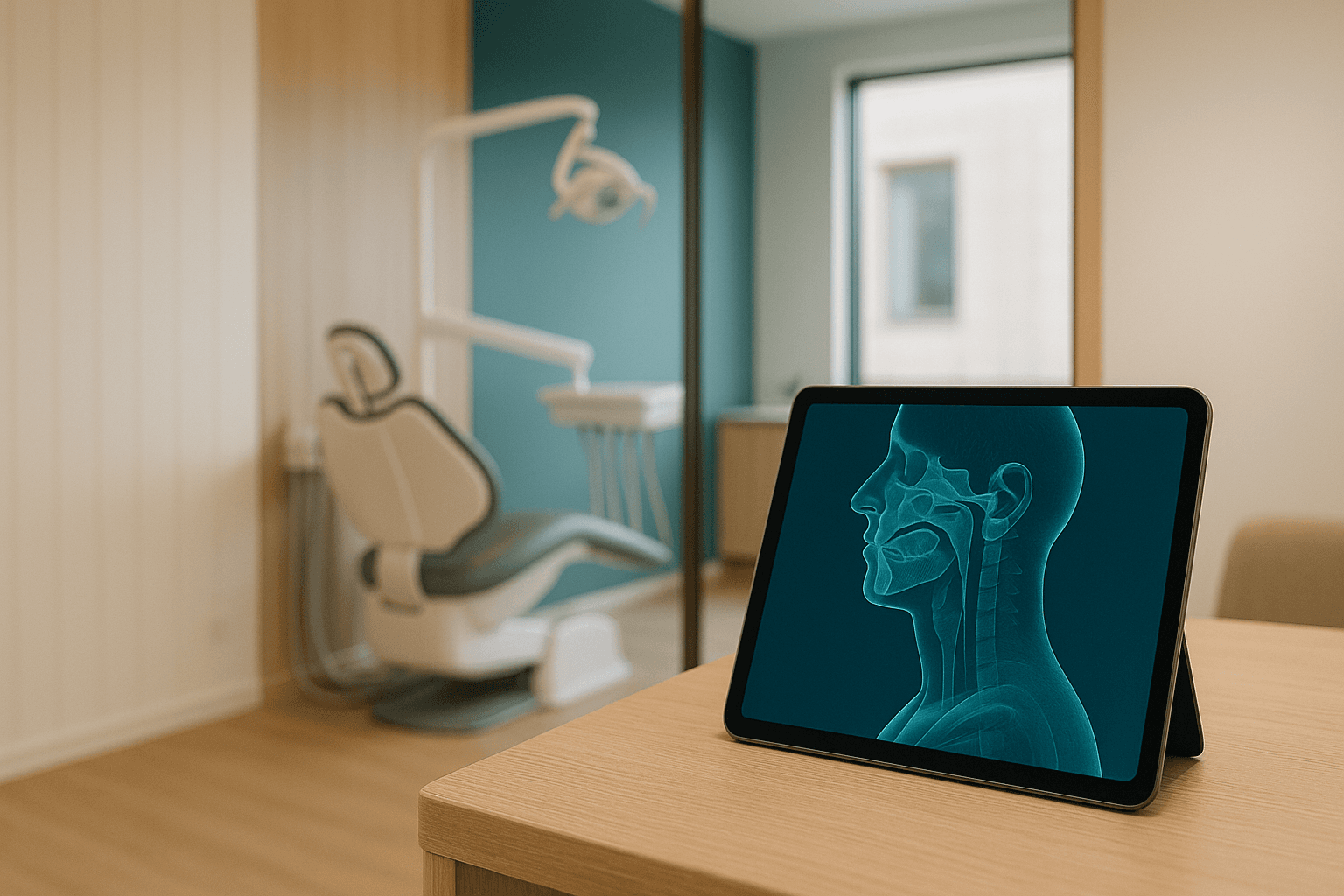
Sit in on a dental study club in 2026 and listen to what the clinicians actually argue about when airway comes up. It is almost never whether the segmentation is accurate.
The argument is about the eleven minutes after the number lands on the screen. The scan was ordered to site a lower right first molar implant, and the software has returned a total pharyngeal volume, a minimum cross-sectional area, and a color-mapped rendering the patient can read upside down from the chair.
An image captured for one purpose, a measurement generated for another, and a patient three feet away waiting for you to say something — that is the operational problem, and it arrives the day you enable the feature. What follows is where the line sits between flagging and diagnosing, and how to build an imaging workflow that stays on the correct side of it.
No. A CBCT is one static image of an awake patient, while obstructive sleep apnea is defined by breathing events during sleep. AI can measure the airway and flag risk; a physician-read sleep study diagnoses it.
What A Vision Model Actually Measures On A CBCT
Modern airway tools segment the pharyngeal air column with a 3D convolutional network — typically a U-Net variant trained on manually labeled volumes — rather than the threshold-and-seed region growing that shipped with earlier planning software. The output is a mesh and a set of numbers, and it helps to know precisely which numbers you are being handed.
Every airway module produces some version of the same measurement set. The values you will see in a report include but are not limited to:
- Total pharyngeal volume. Cubic millimeters of air between a defined superior and inferior boundary, usually the posterior nasal spine plane down to the base of the epiglottis. Where those boundaries sit is a vendor choice, which is the first reason two products disagree on the same scan.
- Minimum cross-sectional area. The smallest axial slice area in square millimeters, reported with its position along the airway axis. This is the metric most closely studied against sleep-disordered breathing, and the one most often quoted out of context.
- Level of the narrowing. Retropalatal, retroglossal, or hypopharyngeal. This matters clinically because it maps to which structures a surgeon or a mandibular advancement device would actually move.
- Shape at the constriction. The anteroposterior-to-lateral ratio at the narrowest slice. A laterally narrowed, elliptical airway behaves differently under negative pressure than a circular one of identical area.
- Adjacent anatomy. Tonsillar and adenoid soft tissue bulk, hyoid position relative to the mandibular plane, and cervical posture. These frequently drive the clinical impression more than the volume number does.
All of these are geometric descriptions of an air column at one instant in a fully awake patient. They are useful, they are reproducible within a single machine and protocol, and none of them measures what happens to that airway at 3 a.m.
Airway modules report total pharyngeal volume in cubic millimeters and minimum cross-sectional area in square millimeters. Both are geometry at one instant, not evidence of what the airway does during sleep.
Why A Narrow Airway Is Not A Diagnosis
Obstructive sleep apnea is defined by events, not by anatomy. Diagnosis rests on an apnea-hypopnea index derived from a sleep study — generally an AHI of 5 or more per hour with symptoms, or 15 or more without them, under the criteria sleep physicians work from.
Anatomy is only one of several traits that determine whether a given airway collapses during sleep. The others — pharyngeal muscle responsiveness, arousal threshold, and ventilatory control loop gain — are invisible to any cone beam scan ever captured.
What's more, the scan is a fragile measurement of even the anatomy it does capture. Airway volume shifts measurably with head and cervical posture, tongue position, mandibular position, upright versus supine positioning, and where in the respiratory cycle the exposure happened to land.
Keep in mind that a patient can present a generous airway on CBCT and still have severe apnea driven by a low arousal threshold or high loop gain. The reverse is just as common: an alarming retropalatal narrowing in a patient whose home sleep test comes back entirely unremarkable.
The Incidental Finding You Just Automated
The practitioner who prescribes a cone beam scan is responsible for interpreting the entire imaged volume, not only the region of interest. That has been the settled position in oral and maxillofacial radiology for well over a decade, and automated airway measurement does not create that duty — it does something harder to manage.
It timestamps it. Your imaging software logged a minimum cross-sectional area at 10:41 with a user ID attached, and the record of what happened in the following ninety days is what a reviewer reads back to you.
Incidental findings on dental CBCT are not rare, and published prevalence estimates vary enormously depending on how broadly the term is defined. Airway narrowing sits among the most frequently identified of them, so automating its detection raises the volume of findings your chart formally acknowledges — sharply and permanently.
Write your flag-and-refer policy before you enable the module, not after the first flag fires on a 41-year-old implant patient with a BMI of 34. The policy is cheap to write in advance and expensive to improvise in the operatory.
The prescribing dentist is responsible for every finding in the imaged volume. Automation timestamps that duty — your software logged a measurement, and the chart now shows what you did about it.
Where The Line Sits Between Flagging And Diagnosing
The clean way to think about scope is to separate three distinct acts: measuring anatomy, screening for risk, and diagnosing disease. A dental practice can do the first two competently, and the third belongs to a physician in effectively every US jurisdiction.
Here is how the common activities sort out, though you should confirm the middle rows against your own state dental practice act and the current state dental board rules on AI in clinical workflows:
| Activity | Dental practice scope | What it actually requires |
|---|---|---|
| Measuring pharyngeal airway volume on a CBCT | In scope as image interpretation | A prescribed scan and documented interpretation of the full volume |
| Telling the patient the airway looks narrowed | In scope as an anatomic finding | Neutral anatomic language, no sleep-disorder label |
| Administering STOP-BANG or the Epworth Sleepiness Scale | In scope as screening | A validated instrument with the score recorded in the chart |
| Stating the patient has OSA or assigning a severity | Out of scope | Physician diagnosis from polysomnography or a home sleep test |
| Ordering the home sleep apnea test | Varies by state; usually physician-ordered | Your board's position plus the state medical practice act |
| Fabricating a mandibular advancement device | In scope after diagnosis | A physician prescription; billed medically under E0486 |
| Titrating or managing CPAP therapy | Out of scope | Sleep medicine physician management |
The boundary tracks the claim you make, not the tool that produced the number. The same minimum cross-sectional area is entirely defensible as a documented anatomic finding and entirely indefensible as a statement that the patient has a sleep disorder.
The practical version of this is a script, and every associate and hygienist in the practice should use the same one. Something close to: “Your scan shows the airway narrowing at the level of your soft palate. That is one of several things associated with breathing problems during sleep, and the only way to know is a sleep study a physician reads — I would like to refer you.”
Note that the script contains a measurement, a qualifier, and a referral, and nothing else. Anything an operator adds beyond those three elements is where the exposure starts, which is also why the incidental-screening disclosure belongs in your AI-assisted informed consent workflow rather than in a hallway conversation.
Compliance Comes Before Model Quality
Before you evaluate a single accuracy metric, settle the data path. A CBCT volume is among the most identifiable artifacts a dental practice holds, and treating it like an ordinary bitewing is the error that turns a screening initiative into a breach investigation.
HIPAA's Safe Harbor list includes full-face photographic images and any comparable images. A full field-of-view maxillofacial CBCT supports surface reconstruction of the patient's face, which makes de-identifying one an expert-determination problem rather than a header-scrubbing exercise.
The DICOM header alone carries PatientName, PatientID, PatientBirthDate, AccessionNumber, InstitutionName, and StationName, plus vendor private tags that differ by manufacturer. Stripping those is table stakes, and it does nothing about the reconstructable face sitting in the voxel data underneath.
Accordingly, the architecture questions that matter here are the boring ones. Here is the shape of a data path that survives an audit:
- An executed BAA with every party that touches the volume. That includes the imaging vendor, the inference provider, and any PACS or cloud archive in the path. Amazon Bedrock is HIPAA-eligible under an AWS BAA; a startup's public inference endpoint frequently is not, and “we do not store your data” is a marketing claim rather than a contract.
- Encryption with keys you control. S3 with SSE-KMS and a customer-managed key, with the key policy held separately from the bucket policy, so revocation is one action instead of a support ticket.
- A private network path. A VPC endpoint or PrivateLink route, so a 200 MB volume never traverses the public internet on its way to inference.
- Data-event logging. CloudTrail data events on the imaging bucket, retained long enough to answer who read which patient's scan and when.
- Minimum necessary at the payload level. Crop to the pharyngeal subvolume before you send anything, which narrows the disclosure and cuts both inference cost and latency.
None of this is exotic, and all of it is the same pattern as any other HIPAA-grade clinical AI deployment in a dental practice. Settle it first, because a model that scores beautifully on a data path you cannot document is not deployable.
Yes. A full field-of-view CBCT supports facial reconstruction, so HIPAA Safe Harbor treats it like a full-face image. Keep the volume under a BAA, encrypted with keys you control.
Whether Your Airway Tool Is A Regulated Device
Vendors will sometimes describe airway measurement as clinical decision support that sits outside FDA device regulation. That framing does not survive contact with the actual policy.
The non-device clinical decision support carve-out explicitly excludes software that analyzes medical images, signals from in vitro diagnostic devices, and patterns acquired from signal acquisition systems. Airway segmentation analyzes a medical image by definition, which places it inside the device framework rather than outside it.
Therefore the question to put to a vendor is short and specific: what is your 510(k) number, what is the cleared indication for use, and does that indication say measurement or does it say detection of sleep-disordered breathing? The distance between those two phrasings is the entire subject of this post.
The FDA's non-device clinical decision support carve-out excludes software that analyzes medical images. Airway segmentation analyzes an image, so ask your vendor for its 510(k) number and cleared indication.
Why Your Airway Numbers Do Not Travel Between Scanners
Cone beam gray values are not calibrated Hounsfield units. They shift with exposure settings, field of view, scatter, beam hardening from restorations, and the reconstruction algorithm, which means the air-to-tissue boundary the segmenter has to find is not consistent from machine to machine.
The downstream effect is that the same patient scanned on a Planmeca ProMax 3D and a Carestream CS 9600 on the same afternoon can produce meaningfully different volumes. Voxel size — typically 0.2 to 0.4 mm in dental protocols — compounds this at the narrowest slice, where a one-voxel boundary shift is a large fractional change in a small area.
This is why published cutoffs make a poor operating threshold. A minimum cross-sectional area figure lifted from a study run on a different scanner, protocol, and segmentation method is a literature reference, not a setting for your fleet.
What works instead is a local baseline. Segment 100 to 200 of your own adult scans, look at your own distribution, set the flag threshold at a percentile of that distribution, and re-check it whenever you change scanners, update firmware, or the vendor pushes a new model.
Pin the model version and record it in the chart alongside the measurement. Version pinning plus routine clinical model drift monitoring is what makes a 2026 measurement interpretable when someone reviews the chart in 2029 — the same discipline that keeps AI radiograph analysis and AI implant planning outputs comparable over time.
What A Defensible Screening Workflow Looks Like
The workflow that holds up is unglamorous and mostly non-technical. Here are the pieces worth building before the first scan runs through the module:
- Write the policy first. One page covering what triggers a flag, who talks to the patient, what words they use, what goes in the chart, and which physicians you refer to by name. Everything else in this list is downstream of that page existing.
- Pair every image flag with a validated instrument. STOP-BANG, the Epworth Sleepiness Scale, Mallampati class, Friedman tonsil grade, neck circumference, and BMI take about four minutes and carry more predictive weight than a volume number standing alone.
- Keep the language anatomic. A narrowed airway at the soft palate is a finding; sleep apnea is a diagnosis you are not licensed to make. The distinction has to survive being repeated by a hygienist on a busy Thursday.
- Refer in writing to a named physician. Send a letter carrying the measurement, the instrument scores, and the anatomic finding to a specific sleep medicine practice, rather than telling the patient to mention it to their doctor.
- Close the loop and track it. Referral sent, study completed, result returned — three fields in Open Dental or Dentrix, reviewed monthly, turn a gesture into a program.
- Get consent for the incidental analysis. The patient consented to imaging for implant planning, so tell them in that consent that the volume will also be screened for findings outside the surgical site.
All of this is a governance workflow with a model attached, and the ordering is deliberate. The measurement is the easy part; the referral loop and the chart language decide whether the program helps patients or quietly accumulates liability — the same lesson that separates useful AI oral lesion screening from an alert nobody acts on.
Record the measurement, the model version, the screening instrument score, the anatomic language used with the patient, and the named physician referral. A flag with no documented referral is the entry you will be asked about.
What This Costs, Honestly
The marginal cost of the inference is close to trivial, because the scan already exists and was already billed — commonly under D0367 for a both-jaws capture and interpretation. Airway modules are usually bundled into CBCT planning software or sold as an add-on, quoted in the low thousands as a perpetual license or a few hundred dollars per month per seat.
The real cost is chair time and follow-through. Budget four to six minutes for the conversation and roughly ten more for the referral letter and documentation, then multiply by the number of adult full-field scans you capture in a month.
On the patient side, the numbers they will ask about are downstream. A home sleep apnea test typically runs a few hundred dollars, an attended in-lab polysomnography study runs well into four figures before insurance, and a custom mandibular advancement device — billed medically under E0486, not through dental benefits — commonly lands between roughly $1,800 and $3,000.
Be aware that the practice captures none of that revenue unless it practices dental sleep medicine, which is exactly why the flagging threshold should be set by someone with no production incentive attached to the outcome. A screening threshold chosen by the person who fabricates the appliance is a threshold nobody wants to defend later.
How Do You Know The Flag Is Working?
Segmentation accuracy is the metric vendors lead with and the least interesting one to an operator. A Dice similarity coefficient in the low-to-mid 0.90s against manual segmentation tells you the mesh is close to a human tracing, and it tells you nothing about whether your referrals are finding disease.
Build the evaluation around outcomes you can actually observe in your own practice. The measurements worth tracking include:
- Test-retest agreement. Volume variance across repeat scans of the same patient on the same machine, which establishes the noise floor beneath every threshold you set.
- Cross-scanner agreement. The same patients imaged on each machine in the fleet, which tells you whether one threshold can serve all your locations or whether each needs its own.
- Referral yield. The share of referred patients who completed a study and returned an AHI of 5 or higher. This is the number that says whether your threshold is calibrated or merely conservative.
- Completion rate. The share of referrals that become completed sleep studies. A flag that produces a 20 percent completion rate is a documentation liability wearing a wellness veneer.
- Missed-case surveillance. Patients who were never flagged and were later diagnosed elsewhere. This is the hardest signal to capture and the most honest one you will get.
Track those five in the same place you track every other model's behavior, and review them on a fixed cadence rather than when someone asks. That is ordinary clinical AI evaluation discipline applied to an imaging feature that happens to touch scope of practice.
Draw The Line Before You Turn It On
Airway measurement off an existing CBCT is one of the highest-leverage screening opportunities in general dentistry, and one of the easiest to implement in a way that generates records nobody wants read aloud. The technical work is a weekend; the policy, the consent language, the referral network, and the audit trail are the actual project.
If you are wiring airway measurement into a CBCT workflow that was built for implant planning or third molar assessment, NexV builds and operates HIPAA-grade clinical AI in production dental environments every week — imaging pipelines, Open Dental and Dentrix integrations, and the audit trails that go with them. Reach out for a working session and we will map your imaging data path end to end, name the scope-of-practice and disclosure failure modes you are about to hit, and leave you with a written flag-versus-diagnose policy your associates can follow on a Tuesday morning.
Definitions And Background Information On AI Airway Screening
The questions below come up in nearly every scoping conversation about airway flagging in a general practice.
Can a dentist tell a patient they have sleep apnea?
No. In effectively every US state, obstructive sleep apnea is a medical diagnosis. A dentist can report a narrowed airway, document a STOP-BANG score, and refer — the diagnosis comes from a physician reading a sleep study.
Is airway measurement software an FDA-regulated device?
Usually yes. The FDA's clinical decision support policy excludes software that analyzes medical images, so airway segmentation off a CBCT falls outside the non-device carve-out. Ask the vendor for its 510(k) number.
Which CBCT airway number predicts sleep apnea?
None reliably on its own. Minimum cross-sectional area correlates with risk across populations, but a single awake, static scan cannot capture the airway collapse that happens during sleep.
Do airway volumes compare across different CBCT scanners?
Poorly. CBCT gray values are not calibrated Hounsfield units, so volume depends on scanner, field of view, voxel size, and segmentation threshold. Build a baseline on your own fleet instead of borrowing published cutoffs.
Does a CBCT volume count as identifiable PHI under HIPAA?
Yes. A full field-of-view scan supports facial reconstruction, which Safe Harbor treats like a full-face image. Keep the volume under a BAA and encrypt it at rest with a customer-managed KMS key.
What should the chart say when the AI flags a narrow airway?
Record the measurement, the model version, the screening instrument score, the neutral anatomic language used with the patient, the named referral, and the date. A flag with no documented referral is the entry you will be asked about.