AI Intraoral Scan Analysis: What Vision Models Can Measure Between Visits, From Tooth Wear to Gingival Recession
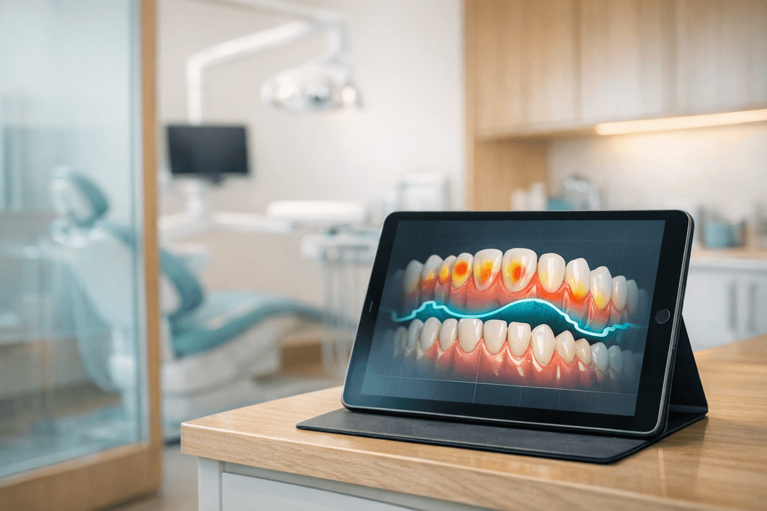
How much enamel did that patient lose on the lower incisors between last year's scan and this one, and how would anyone in the operatory know? For practices that have put an intraoral scanner in every room, the answer increasingly sits in two mesh files that nobody has compared.
That said, comparing them well is an engineering problem before it is a clinical one. This piece separates what vision models can reliably measure across serial intraoral scans from what still belongs on a clinician's desk — and explains where the line moves depending on how the pipeline is built.
Serial intraoral scan analysis aligns two 3D scans of the same arch taken months apart and maps the surface differences. AI can flag wear, tooth movement, and gingival margin shifts; a clinician still decides what they mean.
What An Intraoral Scan Actually Records
An intraoral scan is a triangulated surface mesh, typically exported as STL (geometry only) or PLY and OBJ (geometry plus color). It captures every visible surface the wand reached — enamel, restorations, attached gingiva, and on the maxillary arch, the palatal rugae.
Keep in mind that a scan records surfaces and nothing beneath them. There is no attachment level, no bone height, no pulp, and no subgingival margin, which is why scan analysis complements the AI radiograph analysis a practice already runs rather than replacing it.
What the scan does offer is repeatability. A bitewing is a 2D projection whose geometry shifts with every sensor angle, while two full-arch scans of the same patient can be registered into one coordinate system and subtracted point by point.
Why Serial Comparison Is Harder Than Single-Scan Analysis
Single-scan AI — segmenting teeth, labeling them, spotting a fractured cusp — is largely a solved pattern-recognition task. Serial comparison adds a second problem that most failed pilots underestimate: deciding what did not move, so that everything else can be measured against it.
Registration Is The Whole Game
The standard first step is a best-fit alignment, usually an iterative closest point (ICP) pass across the entire arch. However, a whole-arch best fit quietly assumes that most of the surface stayed still, and in a patient with generalized wear or orthodontic relapse, that assumption is false.
When the reference itself has changed, the alignment distributes the error across every tooth. As a result, real wear on the incisors can be averaged away, and phantom change can appear on molars that never moved.
Practitioners reduce this with stable references rather than whole-arch fits. The options include but are not limited to:
- Palatal rugae registration. The medial rugae on the maxillary arch are widely used in orthodontic superimposition as a relatively stable soft-tissue landmark. They give the upper arch an anchor that does not depend on the teeth being measured.
- Per-tooth local registration. Each tooth is aligned individually on surfaces unlikely to change, such as unrestored buccal and lingual walls. This isolates occlusal wear from whole-tooth movement.
- Multi-stage alignment. A coarse whole-arch fit is followed by a refined fit that excludes regions flagged as changed. The pipeline iterates until the excluded set stabilizes.
The mandibular arch has no equivalent to the rugae, which is why lower-arch comparisons carry wider error bounds. All of this happens before any model looks for wear, and if registration is wrong, every downstream measurement inherits the mistake.
The Noise Floor Sets The Detection Limit
Every scanner, operator, and software version produces a slightly different mesh of the same mouth. Published in vitro studies commonly report full-arch trueness for current-generation scanners in the range of roughly 20 to 60 microns, with wider deviation toward the distal ends of the arch.
Physiological enamel wear, by comparison, is generally estimated at tens of microns per year. In other words, a single annual comparison can sit right at the edge of what the hardware can distinguish from its own repeatability error.
Scanner repeatability error sets the smallest change AI can trust. Measure it with same-day test-retest scans per scanner and operator, then flag only differences that clear that floor by a deliberate margin.
This is why the first deliverable of a serious scan-comparison pipeline is a test-retest study, not a model. Two scans of the same patient, same day, same operator, same scanner, define what zero change looks like — and every flagged finding should clear that floor by a deliberate margin.
What Vision Models Can Measure Reliably Between Visits
Once registration and noise are handled, several measurements become dependable enough to automate as flags. The table below summarizes where each one stands.
| Measurement | Automation readiness | Main error source | Clinician role |
|---|---|---|---|
| Occlusal and incisal wear (volume and depth) | High, over multi-year intervals | Registration bias, scanner noise floor | Diagnose cause: attrition, erosion, or abrasion |
| Tooth position change (drift, extrusion, relapse) | High on the maxillary arch | Lack of a stable mandibular reference | Decide whether movement is clinically relevant |
| Gingival margin migration on facial surfaces | Moderate | Tissue inflammation, retraction, moisture | Confirm with probing; assess etiology |
| Restoration margin change and chipping | Moderate | New restorations between scans | Determine repair versus replacement |
| Shade and surface color change | Low | Lighting, scanner color calibration | Visual exam remains the reference |
Tooth Wear
Wear is the strongest use case because enamel is rigid, the occlusal surface is fully visible, and the change accumulates in one direction. A segmentation network labels each tooth, the pipeline registers each crown locally, and a signed deviation map shows where surface was lost.
From that map, the system can report maximum depth loss, lost volume per tooth, and whether the pattern is localized to the incisal edges or generalized across posterior cusps. Furthermore, comparing the pattern against the opposing arch helps separate tooth-to-tooth attrition from wear that has no matching contact.
Note that the output is a measurement, not a diagnosis. A cupped lesion on a first molar may point toward erosion, yet the decision about dietary history, reflux referral, or a night guard belongs to the dentist.
Tooth Movement
Position change — mesial drift after an extraction, extrusion of an unopposed tooth, or post-orthodontic relapse — is measurable once the arch is anchored. On the maxilla, rugae registration lets the system report translation and rotation per tooth in millimeters and degrees.
Accordingly, scanner manufacturers have built this into their own software, with tools such as iTero TimeLapse and 3Shape's TRIOS Patient Monitoring offering visual comparison between visits. The open question for most groups is whether those vendor views are enough or whether they need structured, per-tooth numbers flowing into the record — mapped to the practice's tooth numbering standard so that FDI and Universal identifiers never get crossed.
Tooth wear and tooth movement are the most automatable serial-scan measurements because enamel is rigid and visible. Gingival changes are measurable but noisier, and color change remains unreliable across visits.
Gingival Recession
Here the line between reliable and unreliable gets thin. A scan can locate the free gingival margin on facial surfaces and measure how far it moved apically relative to a stable point on the crown, such as the incisal edge.
However, true recession is measured from the cementoenamel junction, and the CEJ is often hidden, restored, or simply not resolvable on a mesh. What the system actually reports is gingival margin migration, which is a useful proxy but a different quantity.
Soft tissue also changes for reasons that have nothing to do with attachment loss. Inflammation swells the margin coronally, a retraction cord or cheek retractor deforms it, and pooled saliva can blur the edge the segmentation model needs.
Therefore, a gingival flag should trigger a probing check rather than a chart entry. Practices already running AI periodontal screening on radiographs can treat the scan finding as a second, independent signal that raises priority, not as a replacement for pocket depths.
Operator note: If the pipeline cannot locate the CEJ on a tooth, it should say so explicitly in the output. A margin-migration number presented as a recession number is the kind of quiet mislabel that surfaces later in a peer review or a records request.
What Still Needs Clinician Review
Some determinations stay human regardless of how good the measurement becomes. These include but are not limited to:
- Etiology. Whether wear is attrition, erosion, or abrasion depends on history, diet, habits, and medications that a mesh cannot see. The model can describe the pattern; the clinician names the cause.
- Clinical significance. A 0.4 mm change may be irrelevant on one patient and urgent on another with thin enamel or a history of dentin sensitivity. Thresholds should prompt review, not decide treatment.
- Confounded comparisons. A crown placed between scans, an orthodontic appliance, or a fractured restoration changes the reference geometry. These cases should be routed to a person automatically rather than scored.
- Anything subgingival. Attachment level, bone loss, and subgingival calculus are outside the scan's reach. They still require probing and radiographs.
All of these share one pattern: the scan supplies a precise number, and the decision requires context the scan does not contain. Building the review queue around that distinction keeps the model in its lane.
Clinicians still own etiology, clinical significance, and any comparison confounded by new restorations or appliances. Scan AI should route those cases to review automatically rather than produce a score.
Compliance Comes Before The Model
An intraoral scan linked to a patient is protected health information, and a full-arch color mesh is a large, persistent, identifiable artifact. Before anyone evaluates segmentation accuracy, the data path has to clear the same bar as every other clinical workload — the framework in our piece on HIPAA requirements for clinical AI in dentistry applies directly.
In practice, scans often leave the operatory through the scanner vendor's cloud before they reach any analysis pipeline. Each hop needs a signed BAA, and the practice should know exactly which vendor holds the files, in which region, and for how long.
On the analysis side, the controls are familiar. Here's how they map to a scan-comparison workload:
- Storage. Meshes land in a dedicated S3 bucket encrypted with a customer-managed KMS key, with lifecycle rules aligned to the practice's record-retention policy. Raw files never sit in a shared analytics bucket.
- Network path. Inference runs inside a VPC, with PrivateLink to model endpoints so meshes do not traverse the public internet. The guidance in our Bedrock clinical AI architecture piece covers the endpoint pattern.
- Audit trail. CloudTrail logs every read of a scan object, and the comparison record stores which scan IDs, model version, and registration method produced each finding. When a clinician asks why a flag fired, the answer should be one query away.
Note that the language model — for instance Claude on Bedrock drafting a plain-language summary of the deviation report — never measures anything. It reads structured findings from the geometry pipeline and writes them up, which keeps the numeric claims traceable to deterministic code.
How Do You Know The Comparison Is Working?
A serial-scan system can fail silently in ways a single-image classifier cannot. If the registration drifts, every finding still looks plausible, and nobody notices until a clinician disagrees with a trend line months later.
Build The Validation Suite Around Known Change
The validation suite needs three kinds of cases. First, same-day test-retest pairs establish the noise floor; next, pairs with documented change, such as a crown preparation or a completed orthodontic phase, confirm the system detects what it should; and finally, pairs with a new restoration confirm the system routes confounded cases instead of scoring them.
Our broader framework for clinical AI evaluation applies here, with one addition. Every evaluation result should be stratified by scanner model, because a threshold tuned on one wand will not transfer cleanly to another.
Watch For Drift From The Scanner, Not Just The Model
Scanner firmware and acquisition software update on the vendor's schedule, and an update can change mesh density, smoothing, or color processing. A comparison between a pre-update baseline and a post-update follow-up may show change that is purely an artifact of the software.
Serial-scan pipelines drift when scanner software updates change mesh output. Pin scanner and model versions on every record, rerun test-retest checks after updates, and flag cross-version comparisons.
Accordingly, every scan record should carry the scanner model, firmware version, and acquisition software version, and the pipeline should treat cross-version comparisons as a distinct, reviewed category. The same discipline we describe in model drift monitoring applies, with the input device added to the list of things that can move.
Where Scan Analysis Fits Alongside Radiographs
Most dental AI investment to date has gone into radiographs, and for good reason: caries, periapical pathology, and bone levels live below the surface. Scan analysis adds a different axis — surface change over time — that radiographs capture poorly.
The two work best as complementary signals in the same review queue. A practice running AI caries detection on bitewings and serial scan comparison on the same patients gets interproximal findings from one source and wear, movement, and margin trends from the other.
Furthermore, scan comparisons give the clinician something radiographs rarely do: a visual the patient immediately understands. A color-mapped deviation overlay showing two years of incisal wear is a concrete basis for a night-guard conversation, which is where the operational value tends to show up first.
Overall, the realistic near-term deployment is narrow and useful. Automate registration, measure wear and movement against a known noise floor, flag gingival margin shifts for probing, and route every confounded or cross-version comparison to a person.
Scoping A Serial-Scan Pipeline
If you're evaluating serial intraoral scan analysis across multiple practices and want a second set of eyes on the architecture, the NexV team builds HIPAA-grade clinical AI pipelines on Bedrock that sit alongside the scanner and imaging systems your practices already run. Reach out for a working session — we'll map your scan data path and BAA chain, design the test-retest study that sets your detection thresholds, and leave you with a validation suite you can run before the first flag reaches a clinician.