DICOM and Sensor Interoperability: What Breaks When Clinical AI Reads Radiographs From Three Different Vendors
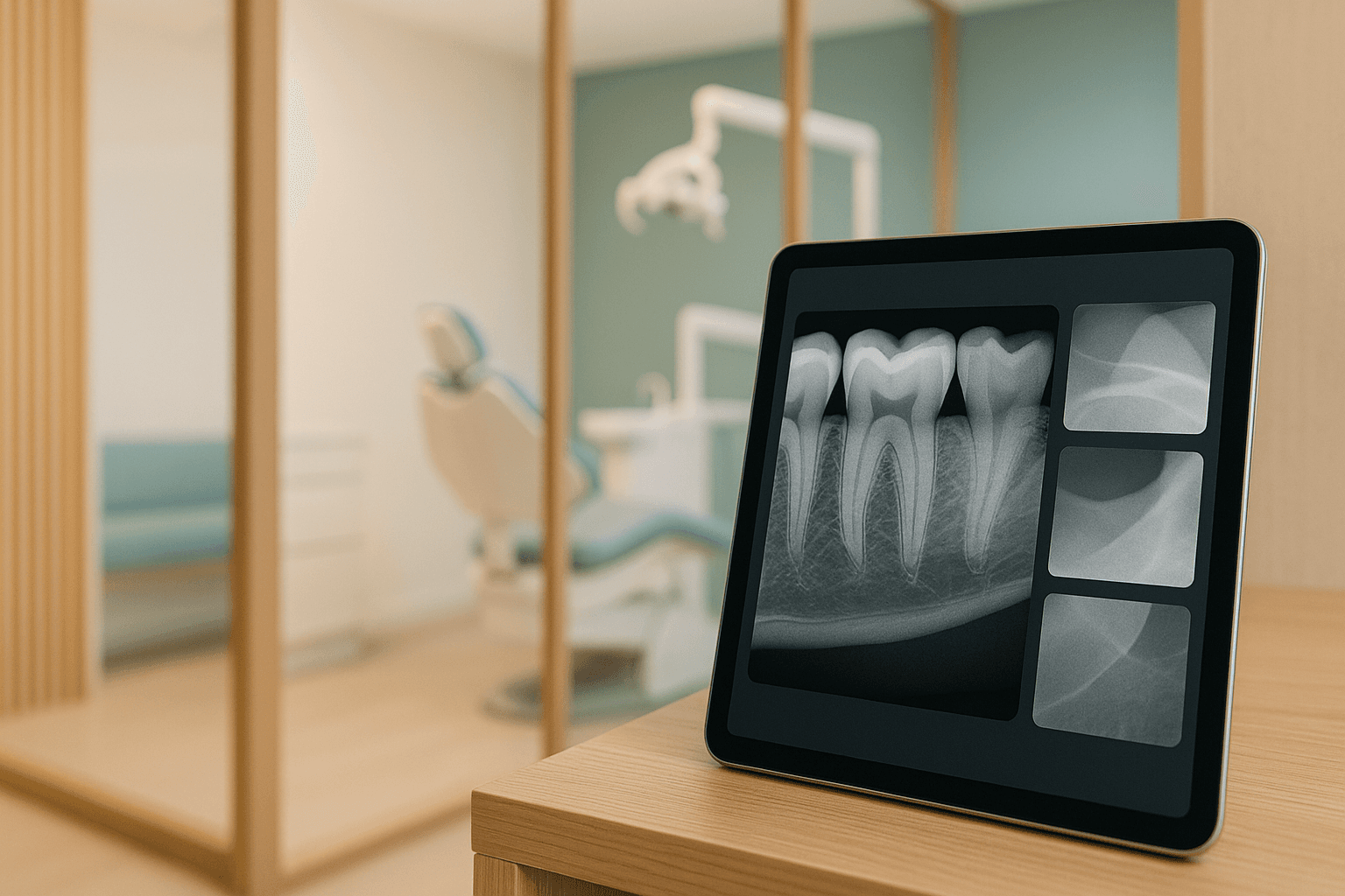
Do you know which of your operatories is exporting 8-bit PNG instead of the sensor's native 16-bit DICOM? If you are running a diagnostic model across more than one imaging vendor, you should — that single export setting moves accuracy more than any hyperparameter you will ever tune.
Most clinical AI post-mortems start at the model and work outward. However, in a practice running three generations of intraoral sensor across six operatories, the damage is usually done upstream — in the capture application, the imaging bridge, and the DICOM header — long before a pixel array reaches inference.
This is the layer that never appears in a vendor demo. The AUC on the slide came from a curated set captured on one sensor line, and the buyer meets the interoperability tax in month four, when a model that read bitewings cleanly in Operatory 2 starts missing interproximal lesions in Operatory 5.
What breaks when clinical AI reads radiographs from multiple sensor vendors?
Bit depth, photometric interpretation, pixel spacing, and vendor tone curves all differ by device. The model sees a different input distribution in every operatory, and accuracy degrades before anyone suspects the pipeline.
Where The Image Pipeline Actually Starts
An intraoral radiograph passes through four owners before your model sees it: the sensor, the vendor capture application, the imaging bridge, and whatever store your practice management system points at. Each hop is an opportunity to discard information irreversibly.
The sensor digitizes 12 to 16 bits of grayscale per pixel. The capture application then applies a proprietary enhancement chain — noise reduction, edge sharpening, and a display curve — and the bridge frequently hands your integration an 8-bit rendering of that processed result rather than the original acquisition.
Note that "DICOM support" on a spec sheet does not mean DICOM fidelity. A file can be a structurally valid DICOM object, pass a conformance validator, and still carry a pre-processed 8-bit pixel array with every tag your preprocessing depends on left empty.
Accordingly, the first artifact worth auditing is the export configuration inside each capture application. That configuration is set per workstation, it changes when a vendor pushes an update, and in most practices it is documented nowhere.
The Bit-Depth Collapse Nobody Logs
Interproximal caries detection lives in low-contrast gradients. A 16-bit acquisition carries 65,536 possible grey levels, an 8-bit export carries 256, and the mapping between them is a vendor curve tuned for a human eye on a specific monitor rather than for a feature extractor.
What is worse, that curve is non-linear and unrecorded. Two sensors can render the same enamel-dentin junction at materially different local contrast, so a model calibrated on one vendor's rendering is reading a systematically different signal on the other.
JPEG compression compounds the loss. Blocking artifacts land exactly where early demineralization lives, and a model trained on lossless input will interpret an 8-by-8 transform edge as a lesion margin without hesitation.
Keep in mind that none of this surfaces in a confidence score. The model returns a well-formed probability on a degraded input, which is precisely why AI caries detection deployments fail quietly instead of loudly.
Why does 8-bit radiograph export hurt caries detection?
It collapses 65,536 grey levels into 256 through an undocumented vendor curve. Low-contrast interproximal signal is compressed away, and the model still returns confident scores on an input that lost the finding.
For Presentation Versus For Processing
DICOM encodes this distinction explicitly, and almost nobody reads it. Projection and intraoral radiographs arrive under one of two storage classes — for presentation, which carries vendor processing and a display curve already applied, or for processing, which carries something much closer to the raw detector read.
A training set that mixes both is a training set containing two different modalities. The pixel statistics differ, the effective noise floor differs, and a model that learned on presentation-grade images will underperform on processing-grade ones even when both came from the same operatory.
For instance, the SOP class UID 1.2.840.10008.5.1.4.1.1.1.3 is digital intraoral for presentation, while 1.2.840.10008.5.1.4.1.1.1.3.1 is the same anatomy for processing. Those two strings differ by two characters and by an entire preprocessing contract.
Therefore the SOP class UID belongs in your ingest gate as a routing decision, not as metadata you log and ignore. Pick one class as canonical, convert or reject the other deliberately, and record which path every stored image took.
What A Three-Vendor Fleet Actually Looks Like
The table below describes an ordinary multi-generation fleet rather than a worst case — three sensor lines installed a decade apart, all feeding one model. Values are illustrative of what these generations typically expose.
| Pipeline attribute | Sensor line A (2013 install) | Sensor line B (2019 install) | Sensor line C (2024 install) |
|---|---|---|---|
| Native pixel depth | 12-bit | 14-bit | 16-bit |
| Detector pixel pitch | Approximately 27 µm | Approximately 19.5 µm | Approximately 15 µm |
| What the bridge exports | 8-bit JPEG via a scanner interface | 8-bit PNG or DICOM for presentation | 16-bit DICOM for processing |
| Imager Pixel Spacing (0018,1164) | Absent | Present, profile-dependent | Present and populated |
| Photometric Interpretation | MONOCHROME2 | MONOCHROME1 on some export paths | MONOCHROME2 |
| Enhancement chain | Baked in, non-reversible | Baked in, varies by user profile | Optional, off for processing output |
| Device Serial Number (0018,1000) | Absent | Present | Present |
Read the export row first. Two of these three paths destroy information that no downstream normalization can recover, which means the fix is a configuration change in the operatory rather than a transform in your pipeline.
The Tags That Decide Whether Your Model Is Looking At The Right Image
A DICOM header is a contract, and a multi-vendor fleet is a set of vendors honoring different parts of it. The tags that most often break a radiograph pipeline include but are not limited to:
- Photometric Interpretation (0028,0004). MONOCHROME1 means the lowest stored value displays as white, while MONOCHROME2 means it displays as black. Ignore this tag and part of your fleet arrives tonally inverted, which a vision model reads as an entirely different image.
- Imager Pixel Spacing (0018,1164). This is the physical size of a detector element and the only reliable way to convert pixels into millimeters on a projection radiograph. Intraoral exports omit it frequently.
- Rescale Slope and Intercept (0028,1053 and 0028,1052). Applied in the wrong order or skipped entirely, stored values stop mapping to the intensities the model was trained on.
- Window Center and Width (0028,1050 and 0028,1051). These describe a display preference, not the image. Baking them into the tensor is one of the most common ways a team accidentally trains on a monitor setting.
- Modality (0008,0060). Intraoral should be IO, panoramic PX, and CBCT CT, but a generic OT value and a miscoded DX appear often enough that modality-based routing needs an explicit fallback.
- Manufacturer, Model Name, and Device Serial Number (0008,0070, 0008,1090, and 0018,1000). Without these you cannot stratify accuracy by device, which means a per-operatory regression is invisible to you by construction.
- Presentation LUT Shape (2050,0020). An INVERSE value flips the intended rendering, and de-identification tooling strips this tag more often than it preserves it.
All of these are cheap to validate at ingest and expensive to discover in production. A tag contract with required fields, allowed values, and explicit rejection turns a silent accuracy problem into a loud queue problem, which is the trade you want every time.
Which DICOM tag most often breaks AI radiograph pipelines?
Photometric Interpretation (0028,0004). MONOCHROME1 and MONOCHROME2 invert the grayscale relationship, so ignoring it feeds tonally inverted images to a model never trained on them.
Pixel Spacing Decides Whether Millimeter Outputs Mean Anything
Periodontal bone loss, root length, and lesion depth are metric claims. A model reporting 3.1 mm of attachment loss is doing arithmetic on pixel spacing, and if that value was missing and silently defaulted, the number is fiction wearing a unit.
The scale problem reaches pure classifiers too. A pipeline that resizes every input to 512 by 512 without normalizing physical scale presents the same molar at meaningfully different apparent sizes across sensors at 15 µm and 27 µm pitch — close to a 1.8-times difference in millimeters per pixel.
Never default-fill a missing spatial tag. Route that study to a dead-letter queue, surface it as a device configuration defect, and fix it at the operatory, because a plausible constant is the most dangerous value you can write into an imaging pipeline.
Our AI periodontal screening work treats calibrated spacing as an admission requirement rather than a nice-to-have. Radiographic bone-loss output stays suppressed on any study that cannot prove its scale.
Does pixel spacing matter if the model only classifies?
Yes. Fixed-size resizing without scale normalization changes the apparent size of anatomy across sensors with different pixel pitch, shifting the input distribution even when no measurement is reported.
Calibration Drift Turns One Operatory Into Its Own Failure Mode
Sensors age. Scintillator efficiency declines, dead pixels accumulate, and the stored gain and offset calibration gradually stops matching the detector it was measured against.
The visible result is a gradient, a vignette, or a line artifact that a human reader compensates for without noticing. A model does not compensate — it treats the artifact as signal, and it does so consistently enough to resemble a real clinical pattern confined to one operatory.
Exposure technique adds a second axis of drift. kVp, exposure time, and holder geometry vary by operator, so two operatories running identical hardware still produce different density distributions and different rates of cone-cut and foreshortening.
The operational rule: any sensor whose last gain and dead-pixel calibration is older than your monitoring interval is out of specification for AI inference, whether or not its images still look fine to a clinician.
Accordingly, calibration date belongs in the same provenance record as model version. When per-device accuracy dips, the first question is whether that sensor was recalibrated, and you cannot answer it retroactively if you never stored it.
This is the same failure surface described in our work on model drift monitoring, arriving through hardware rather than through data. The weights are unchanged; the world in front of the detector is not.
What is sensor calibration drift in dental imaging?
It is the slow divergence between a detector's stored gain, offset, and dead-pixel map and its current physical behavior. It produces gradients and artifacts that a model reads as clinical signal.
DICOM Headers Are PHI, So Normalization Happens Inside The Compliance Boundary
Before this is an accuracy project it is a compliance project. A DICOM header carries patient name, patient identifier, birth date, study date, and referring provider, and intraoral studies sometimes carry burned-in annotation in the pixel data itself.
That makes every normalization hop — bridge export, tag rewrite, resize, tensor cache — an in-scope transformation of protected health information, requiring a signed BAA with whoever operates it. The normalizer therefore belongs inside your own VPC with KMS encryption at rest, CloudTrail logging on every object read, and PrivateLink to the inference endpoint, not in a convenient third-party conversion utility.
De-identification has to be tag-aware rather than blanket. A scrubber run at profile strength will remove Imager Pixel Spacing, Device Serial Number, and private-group enhancement parameters — the exact tags your pipeline depends on — so the de-identification allow-list and the model's tag contract must be written together.
The full architecture requirements appear in our breakdown of HIPAA-compliant clinical AI in dental practices, and the isolation model for inference itself in running clinical AI on Amazon Bedrock. Both apply to images exactly as they apply to text.
The Normalization Layer That Recovers Most Of The Loss
Once the compliance boundary is settled, the fix is an ingest layer that refuses to guess. In practice it carries six responsibilities:
- Enforce a tag contract. Required tags, allowed values, and explicit rejection on violation, evaluated before any pixel work happens.
- Prefer the least-processed source. Route for-processing images wherever they exist, and record every case where a for-presentation image was accepted as a fallback.
- Canonicalize deliberately. One grayscale convention, one bit depth, one linearization step, and one normalization to millimeters per pixel, all versioned as code rather than configured per site.
- Version the preprocessing. Preprocessing drift is as real as model drift, so the transform version gets pinned and stored beside the model version for every inference.
- Stamp provenance on every study. Manufacturer, model name, serial number, SOP class, calibration date, and transform version, written immutably next to the result.
- Dead-letter, never default. Studies failing the contract go to a queue a human works, with the operatory and workstation named in the alert rather than buried in a log.
Note that this layer costs latency, and chairside tolerance for latency is thin. Budget the normalization pass explicitly against the numbers in our chairside AI latency analysis instead of discovering it at the chair.
All of this adds up to a pipeline where a bad image produces an alert instead of a diagnosis. That is the only version of this system that is safe to run without a human watching every study.
How do you normalize radiographs from multiple DICOM vendors?
Enforce a tag contract at ingest, prefer for-processing sources, canonicalize grayscale and millimeters per pixel, version the transform, and dead-letter failures instead of default-filling missing tags.
Proving It Works Before It Reads A Live Radiograph
A single fleet-wide accuracy number hides exactly the failure this post describes. Stratify every metric by device model, by operatory, and by SOP class, then hold each stratum to the same threshold rather than holding the average to it.
Next, run shadow mode per device rather than per practice. A model can be production-ready on the 2024 sensors and unfit on the 2013 ones, and a practice-level rollout decision has no way to express that difference.
When accuracy does move, the triage table below covers most of what you will find. Each row is a question about provenance rather than a question about the model.
| Symptom | Usual cause | Where to look first |
|---|---|---|
| Strong in some operatories, weak in others | Export configuration or sensor generation difference | Capture-app export settings; manufacturer and model tags per operatory |
| Findings inverted or wildly over-flagged | MONOCHROME1 processed as MONOCHROME2 | Photometric Interpretation and Presentation LUT Shape |
| Millimeter outputs plausible but wrong | Missing pixel spacing that was default-filled | Imager Pixel Spacing presence rate per device serial |
| Accuracy dropped after an archive re-export | Historical images re-rendered through a current display curve | Migration job logs; SOP class distribution by ingest date |
| Gradual decline in one operatory only | Sensor calibration drift | Gain and dead-pixel calibration date for that serial number |
| Fleet-wide shift with no deployment | Capture-application update changed the enhancement chain | Capture software version history per workstation |
Every row here is diagnosable in an afternoon when provenance was stored, and effectively undiagnosable when it was not. That asymmetry is the whole argument for the provenance record, and it is the same argument we make for clinical AI evals in general.
What A Migration Window Does To All Of This
Migrations are where image pipelines break at scale, because a migration touches the archive rather than the stream. A cutover that re-exports fifteen years of radiographs through today's imaging bridge can rewrite the tonal character of every historical study in a single weekend job.
If that archive is also your ground-truth set, the evaluation baseline just moved without a code change. Freeze and hash the evaluation set before the migration window opens, then re-score against the frozen copy afterward to measure what the re-export actually did.
What is more, tooth numbering conventions travel badly. A source system storing Universal numbering from 1 to 32 feeding a model that assumes FDI two-digit notation produces confidently mislabeled findings — a mapping defect rather than a vision defect, and one covered in our work on AI dental charting.
The sequencing questions are the same ones worked through in Dentrix Cloud migration planning and chart consolidation. Treat the imaging bridge as a first-class migration workstream instead of an afterthought that trails the financial cutover.
Frequently Asked Questions About DICOM And Sensor Interoperability
These questions come up in nearly every scoping conversation about multi-vendor imaging.
Does DICOM support guarantee interoperability between imaging vendors?
No. A file can be valid DICOM and still carry an 8-bit processed pixel array with pixel spacing, photometric interpretation, and device tags left empty, so fidelity has to be verified per device.
Should you retrain per sensor vendor or normalize the inputs?
Normalize first. Per-vendor models multiply your validation, monitoring, and pinning burden by the size of the fleet, while a versioned normalization layer holds one model to one input contract.
How much accuracy is lost when a model reads a new sensor vendor?
External validation on images from an unseen device commonly costs ten to twenty points of sensitivity. Measure on your own captures per device before trusting any vendor-published number.
How often should intraoral sensors be recalibrated for AI use?
At least as often as your drift monitoring interval, and on the manufacturer's schedule at minimum. Store the calibration date as provenance so a per-device accuracy dip can be checked against it.
Can 8-bit radiograph exports be converted back to diagnostic-grade input?
No. Bit-depth reduction and a baked-in display curve are lossy and non-invertible, so the only real fix is changing the export configuration at the capture application.
Do DICOM headers require de-identification before model training?
Yes, but tag-aware de-identification. Blanket scrubbing removes pixel spacing, device serial, and private enhancement tags, so write the allow-list and the model tag contract together.
Scoping A Multi-Vendor Radiograph Pipeline
The image pipeline is the cheapest place to buy accuracy and the last place most teams look. A model that reads three vendors well is usually the same model sitting behind an ingest layer that refuses to guess.
If you are scoping diagnostic imaging across more than one sensor vendor and want a second set of eyes before it reads a live radiograph, the team at NexV builds and operates HIPAA-grade clinical AI inside dental practices every week. Reach out for a working session — we will audit the export path and DICOM tag coverage of every device in your fleet, define the per-device and per-SOP-class metrics your evaluation is currently missing, and hand back a normalization and shadow-mode plan your clinicians can sign off on.